Whole Slide Image Viewer
CMU-1-Small-Region |
CMU-2-Large-Region |
CMU-3-Large-Region |
Guppy-Sagittal-Plane |
Rabbit-Tongue |
Cat-Claw
Info
Close
Project Info
This web based whole slide image (WSI) viewer implements an open source workflow to process and display digital microscopy images captured in DICOM or Aperio SVS formats. Deep Zoom and OpenSeaDragon have been leveraged to provide high performance panning and magnification of images over 2 gigabytes in size while consuming minimal bandwidth and compute.
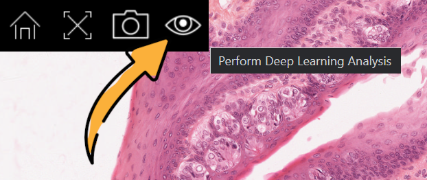
Multimodal deep learning models including LLaVA have been integrated into the viewer to provide image analysis on-demand. Press the analysis button as pictured below to submit the active viewport for analysis.
Home: HTTP://BRYCENICHOLS.NET
Cookie Policy: HTTP://BRYCENICHOLS.NET/COOKIES